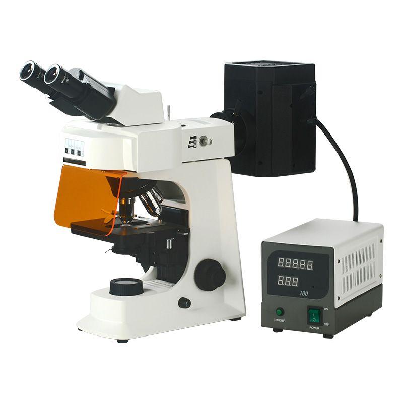
What Measures Can Be Used to Quantify Fluorescence Microscopy Data?

There are many ways to quantify fluorescence microscopy data. Some common methods include:
l Image analysis software: Fluorescence microscopy images can be examined using a variety of software programs. These computer programs can be used to calculate the fluorescent signal's area, intensity, and shape.
l Fluorescence lifetime imaging microscopy (FLIM): FLIM is a method that measures the fluorescent signal's lifespan under a microscope. The fluorescent dye can be recognized and its concentration can be measured using the lifespan of the fluorescent signal.
l Fluorescence spectroscopy: Fluorescence spectroscopy is a method for determining the emission spectra of a fluorescent dye using a spectrometer. The fluorescent dye can be recognized and its concentration can be measured using the emission spectrum.
l Image cytometry: Using a microscope to picture cells and software to analyze the images is a technique known as image cytometry. Quantifying the fluorescent signal in individuals or populations of cells is possible with image cytometry.
The most effective technique for measuring data from fluorescence microscopy will vary depending on the application. For instance, image analysis software would be used if you wanted to measure the strength of the fluorescent signal in certain cells. Image cytometry would be used if you wanted to determine the amount of a fluorescent dye present in a population of cells.
- Art
- Causes
- Crafts
- Dance
- Drinks
- Film
- Fitness
- Food
- Juegos
- Gardening
- Health
- Inicio
- Literature
- Music
- Networking
- Otro
- Party
- Religion
- Shopping
- Sports
- Theater
- Wellness


