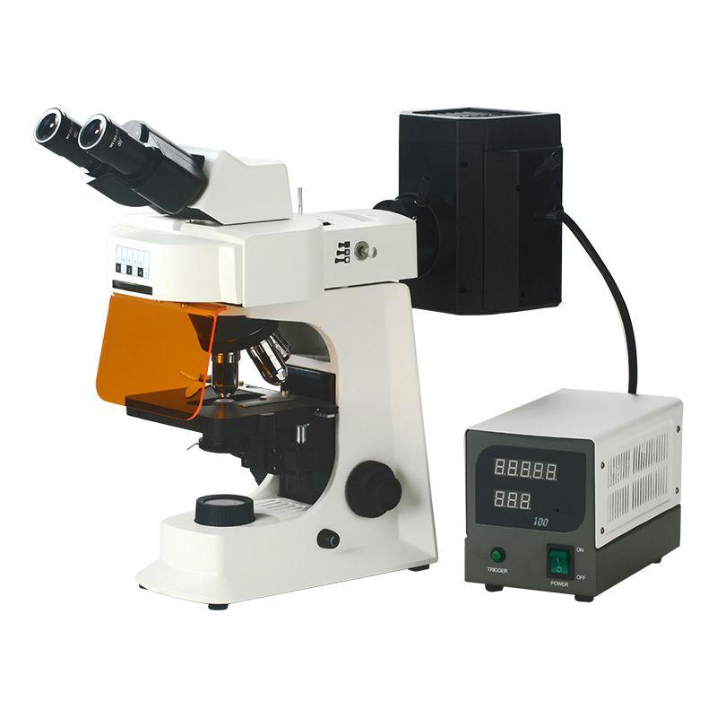
What Are the Advantages of Fluorescence Microscope?

Fluorescence microscopes offer several significant advantages over traditional biological microscopes, making them a powerful tool in various fields of biology and medicine. Here's a closer look at some key benefits:
High Specificity
l Targeted Visualization: Unlike traditional microscopes that rely on general staining techniques, fluorescence microscope allows researchers to target and visualize specific structures within a cell or sample. This is achieved by using fluorescent molecules called fluorophores that bind to specific proteins, organelles, or other cellular components of interest. When excited by light, these fluorophores emit their own light, creating a bright image against a dark background. This targeted approach allows scientists to focus on specific aspects of a cell, eliminating interference from other cellular components.
l Multiple Labeling: Fluorescence microscope offers the ability to use multiple fluorophores simultaneously, each with a distinct emission color. This allows researchers to visualize several different structures within a single cell, providing a more comprehensive picture of cellular processes or interactions between different molecules.
High Sensitivity
l Detecting the Faint: Fluorescence microscope is incredibly sensitive. It can detect very small amounts of fluorophores, making it ideal for observing low-abundance molecules within a cell. This is particularly beneficial for studying proteins that are present in very small quantities or for identifying rare pathogens within a sample.
Enhanced Detail and Information:
l Clearer Images: Because fluorescence microscope uses targeted labeling and eliminates background noise from unstained structures, it can produce clearer and more detailed images compared to traditional brightfield microscope. This allows for better visualization of cellular structures and finer details within a specimen.
Live-Cell Imaging:
l Observing Dynamics: Certain fluorescence microscope techniques can be used for live-cell imaging. By tagging specific molecules with fluorophores, researchers can observe their movement, localization, and interactions within living cells in real-time. This provides valuable insights into cellular processes and dynamics that wouldn't be possible with fixed samples.
While fluorescence microscope offers a powerful toolset, it's important to consider some limitations, such as potential photobleaching (fluorophores losing their fluorescence over time) and autofluorescence (natural fluorescence from cellular components). However, the advantages of high specificity, sensitivity, and detailed information make fluorescence microscopy a cornerstone technique in biological research.

- Art
- Causes
- Crafts
- Dance
- Drinks
- Film
- Fitness
- Food
- Jeux
- Gardening
- Health
- Domicile
- Literature
- Music
- Networking
- Autre
- Party
- Religion
- Shopping
- Sports
- Theater
- Wellness


